CME: Mirror Therapy for Phantom Limb Pain and Stroke
- FibonacciMD

- Aug 14, 2025
- 10 min read
Updated: Aug 19, 2025
In this Continuing Medical Education article, discover how mirror therapy can ease phantom limb pain and aid stroke recovery through brain reprogramming.
Read Article, Take Test, Get FREE 🎓CME Certificate with Valid Email
We’ll send you occasional updates. Your email stays private—never sold or shared. 😇

By Stuart M. Caplen, MD
After a limb amputation, a patient may feel as if the limb is still attached. This can be a source of severe pain and is known as phantom limb syndrome. It occurs in up to 90% of people with limb loss.[1] Mirror therapy has been shown to decrease the false perception of having the limb attached, as well as reducing the subsequent pain that can occur. The process by which mirror therapy is theorized to work illustrates both the complexity and plasticity of the human brain.
What is Phantom Limb Pain?
Phantom limb syndrome patients can experience movement, pain, and muscle spasms that feel as if they are coming from the amputated limb. Perceived movements can be complex, such as waving goodbye, or the phantom limb may feel as if it is frozen in place. Theoretically, it is postulated that the motor cortex does not “realize” that the limb is missing and continues to try to move it. The motor cortex of the brain continues to send messages to the muscles in the amputated extremity that it still “believes” are there. These signals can get amplified when the motor cortex does not receive feedback that the movement has occurred.[2] Movement directives for the limbs are monitored by the parietal lobes, which are involved with body image. The parietal lobe may be where an amputee’s perception of movement in the phantom limb is generated. It may be due to the contradictory processes of the brain “thinking” the limb is still attached and sending out increasingly amplified messages to the limb, while the visual system “sees” there is no limb, which leads to phantom limb pain.[2] Another theory is that pathological remapping of the brain after an amputation may lead to chaotic, abnormal outputs that lead to a perception of pain in the phantom limb.[3]
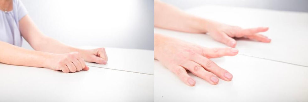
Mirror therapy appears to work by creating the illusion that both limbs are moving normally, which reduces the conflict between proprioceptive and visual inputs (the feeling that the limb is there and moving, but not visualizing the limb or seeing it move) and reprograms the brain. In mirror therapy, the person puts the uninjured limb in either an open box with a mirror in it, designed for that purpose, or simply uses a mirror placed on a table or floor. Moving the unaffected arm or leg and viewing it in the mirror creates the illusion that both limbs are present and moving, thus reducing the incompatibility of the visual and proprioceptive brain inputs, and potentially decreasing troubling phantom limb symptoms.[2]
What Observation Led to Mirror Therapy?
Interestingly, patients who had paralysis in a limb before amputation typically feel as if the phantom limb is also paralyzed (called learned paralysis), while those whose limb was not paralyzed can “feel” the limb move for some time until eventually, due to lack of sensory feedback, they may lose that ability. It was this observation that led to the creation of mirror therapy.[2]
In patients who previously had paralysis of a limb prior to its amputation, each time the message to use the paretic limb went from the motor cortex to the arm, the brain received visual feedback that the arm was not moving. This appears to get incorporated into the neural circuitry of the parietal lobes so that the brain “learns” that the paralyzed arm is fixed in that position. After an amputation of a paralyzed limb, the brain already “knows” the arm does not move, and the phantom limb does not move either. The concept that the brain can “learn” and be “rewired” by the use of visual stimuli to accept that the paretic limb was paralyzed, was considered central to the concept of mirror therapy by its creators. Their theory was that as the brain had demonstrated that it could be “rewired”, if they created a visual message to the brain that a task the motor cortex wanted to be performed by a phantom limb was in fact performed, it might possibly reduce phantom limb pain.[2]
To accomplish that, a mirror box was used to create the illusion that the amputated limb was actually moving, using a mirror image of the intact limb. This was done to provide visual feedback in hopes of reprogramming the brain. In one of Ramachandran’s and Rogers-Ramachandran’s early experiments, a patient who had a left arm amputation nine years earlier and could not “move” his phantom left limb, initially tried to move both his right arm and phantom left arm while in the mirror box with his eyes closed. While his normal right arm moved, his left arm felt “frozen as if in a cement block.” However, when he opened his eyes and viewed the mirror image of what now appeared to be a normal left arm, he experienced vivid sensations of movement in his phantom limb. The authors felt that the ability to feel movement in a previously frozen phantom arm, after visualizing a normal mirror image, implied that new neural pathways could be created in the adult brain. It also implied that areas of the brain concerned with vision and proprioception must interact to a great extent, with visual feedback exerting a modulating effect.[2,3]
Does Mirror Therapy Work ?
In 1996, Ramachandran and Rogers-Ramachandran published an article titled “Synesthesia in Phantom Limbs Induced with Mirrors”. (Synesthesia is the production of a sensation to one sense or part of the body by stimulation of another sense or part of the body.) They studied ten subjects who had phantom limb syndrome after an amputation and felt either pain or spasms in their absent limbs. They then had them use a mirror box, which presented the visual illusion of having two normal limbs. In six of the subjects, moving the normal limb, which when seen in the mirror looked like the subject had two normal limbs, caused the subjects to “feel” movement in the phantom arm. Four out of five patients who had experienced recurrent painful clenching spasms of their phantom hands experienced relief from those spasms when the normal hand was opened. and they could see that the mirror image hand was also open. It was not possible for those subjects to get pain relief without viewing the mirror image of the normal, unclenched hand in place of the amputated limb. In total, eight of the ten subjects were able to get pain relief from the use of mirror therapy. In three patients, touching the normal hand, which when viewed as a mirror image, induced precisely localized touch sensations in the phantom hand. In another subject who had symptoms for ten years, three hours of visual input using a mirror box over three weeks permanently altered his body image. His phantom arm disappeared, and all he felt after therapy was part of the palm and fingers dangling from his shoulder. He also eliminated his chronic phantom elbow pain, and the therapy allowed him to “move” his phantom fingers rather than feeling them painfully clenched and fixed in position. In another experiment, they asked subjects to attempt to place their phantom hand in the mirror box palm down. The examiner then put his gloved hand in the box palm up, which could be seen in the mirror. When the examiner flexed his fingers, the subjects complained that their phantom hand was painful and felt the fingers were being hyperextended into anatomically impossible positions.[2]
In support of their theory of brain neuroplasticity, Ramachandran and Altschuler discussed a case where three weeks after a traumatic amputation, a patient felt sensation in his phantom hand when certain areas of his contralateral face were touched. In the brain architecture, the motor and sensory areas of the hand are located next to the face. It has been shown that in some patients with phantom limbs there is an extension of the brain’s face area into the hand area, which implies neuroplasticity.[3]
A different, well-known example of brain plasticity is that some blind musicians process auditory inputs in both the auditory and optical brain cortices.[4]

More Mirror Therapy Studies
Ramachandran’s work was repeated by others, and in one experiment by Chan et al., 22 subjects with leg amputations and phantom limb pain were separated into three groups. One group was treated with a mirror box and attempted to use both limbs while viewing the mirror; one group used a mirror box with the mirror covered up; and one group used just mental imagery to pretend to move both limbs. They performed these tasks for 15 minutes a day for four weeks. At the end of that time, 100% of the mirror-treated patients reported a decrease in pain versus 17% in the covered mirror group and 33% in the mental-visualization group. After the initial experimental endpoint, 89% of those in the covered mirror and mental-visualization groups who switched to mirror therapy reported decreased pain after a second 4-week period.
The authors stated that, “Pain relief associated with mirror therapy may be due to the activation of neurons in the hemisphere of the brain that is contralateral to the amputated limb. These neurons fire when a person either performs an action or observes another person performing an action. Alternatively, visual input of what appears to be movement of the amputated limb might reduce the activity of systems that perceive protopathic pain.”*[1]
* (Protopathic pain is a type of sensory perception characterized by a generalized, non-discriminating response to stimuli like pain or temperature.)
Ramadugu et al. studied 60 amputees with mirror therapy using a control group where the mirror was covered up. They reported significant decreases in pain in the mirror therapy group versus the control group. When the control group was crossed over to mirror therapy, they also had significantly decreased phantom limb pain up to 12 weeks after starting therapy.[5]
Finn et al. tested 15 male upper extremity amputees and compared mirror therapy, to either covered mirrors or mental-visualization therapy, as the control groups. Therapy was performed five days a week for four weeks. 89% of subjects in the mirror therapy group had a significant decrease in pain scores. The mean amount of time spent daily by the group experiencing pain also significantly decreased over the controls. The control groups did not experience a significant diminishment of pain, nor a decreased overall time experiencing pain. After four weeks, five out of six of the control group subjects switched over to mirror therapy and all experienced decreased pain and reduced daily time spent in pain.[6]
Another study by Yildirim and Kanan reported that mirror therapy significantly decreased phantom limb pain in 15 amputees, with pain decreasing each week over four weeks of treatment.[7]
A meta-analysis of mirror therapy for phantom limb pain reported that there was a significant decrease in phantom limb pain after one month with mirror therapy, with those subjects who had the pain for more than a year getting the most benefit. There was no evidence of long-term benefit of mirror therapy, but the authors stated this might be due to limited data from the literature available at the time they authored the article.[8]
A systematic review found that mirror therapy worked to reduce phantom limb pain, but there was limited scientific data supporting its effectiveness.[9] Another systematic review reported that the level of evidence was insufficient based on the data reviewed to recommend mirror therapy, and more studies were needed.[10]
Mirror Therapy for Stroke Victims
In 1999, Altschuler et al. performed a pilot study of mirror therapy on nine stroke patients with post-stroke hemiparesis. Among these subjects, three showed moderate recovery, three exhibited mild recovery, and three experienced no change. Their theory was that there is a temporary interruption of brain signals after a stroke, which leads to a form of “learned paralysis” similar to that seen in phantom limb. This may persist even after the post-stroke swelling and edema subside, at which point mirror therapy might help recovery.[3,11]
Movement, as seen using mirrors, has been found to add additional activation of the hemisphere contralateral to the affected limb, which might also help stroke victims. The mirror imagery is thought to increase cortico-muscular excitability, which might directly improve motor functioning. It is also hypothesized that mirror therapy may normalize central sensory processing by providing a visual image of the affected limb as having normal movement, which may help reduce pain after a stroke.[12]
A Cochrane review looked at the use of mirror therapy to improve limb motor function after a stroke. They included 62 studies with a total of 1,982 subjects who had suffered a stroke and concluded that there was moderate‐quality evidence that mirror therapy has a significant positive effect on motor function and motor impairment and may improve the ability to do activities of daily living. There was low-quality evidence for a significant positive effect on reducing pain. They also reported there was no clear effect on improvement of visuospatial neglect symptoms (failing to notice, respond to, or report stimuli on the side opposite their brain lesion). The improvement in pain was noted mostly in individuals with complex regional pain syndrome. The authors noted that there were limitations on the use of data from the current literature on mirror therapy after stroke due to small sample sizes and lack of reporting of methodological details, resulting in uncertain evidence quality.[12]
Summary
An intuitive leap led to the creation of mirror therapy, based on the observation that previously paralyzed phantom limb patients did not feel movement, but those who had normal functioning limbs before amputation could feel the missing limb still move. This “learned paralysis” indicated the brain could be “rewired.” Ramachandran thought that phantom limb pain was due to discoordination between the motor cortex and visual inputs, and the brain could be “tricked” into thinking the limb was still there by use of a mirror. Using a mirror to create normalized limb visual input has been shown to reduce phantom limb symptoms in many patients and should be considered when recommending therapy for patients who suffer from this condition.
Mirror therapy may also be helpful in rehabilitating stroke victims who have limb paresis.
The fact that this relatively simple, but ingenious therapy appears to work is an indication of both the complexity and the plasticity of the human brain.
Note: There is also some literature evidence, not discussed in this article, that motor imagery (where patients imagine the limb is moving normally) and virtual reality visual feedback of normal limb movement (similar to mirror therapy) may also be helpful in some stroke or phantom limb patients.[1,9,13]
Author’s Note: Thank you to Dr. Theodor Feigelman for editing this article.
✅ Earn Free CME Credit for Reading This Article
Eligible for 0.5 PRA Category 1 Credit
Click the button below to take a short quiz.
A valid email is required to send your certificate.
We’ll send you occasional updates. Your email stays private—never sold or shared. 😇
Earn CME Credit NowBUTBU