Search Results
Search this site
376 results found with an empty search
- Thyroid Nodules: Clinical Guidelines, Risk Factors, and Diagnostic Work-up
Understanding Thyroid Nodule Malignancy Risks and the Gold Standard for Diagnosis FibonacciMD Compendium Otolaryngology Term The Thyroid Nodule Photomicrograph showing histology of a benign thyroid nodule in a patient ith multinodular goiter. Follicles of varying size are seen, many filled with colloid material. By Michael Bergstein, MD Definition of Thyroid Nodule Thyroid nodules are soft tissues lesions found within the thyroid gland. They are quite common, and can be found in both adult and pediatric patients. Approximately 95% of thyroid nodules are benign. The risk of malignant disease increases in young populations (< 20) and older populations (women > 40 and men > 50). The vast majority of malignant thyroid nodules are papillary thyroid carcinoma. Thyroid nodules can also be present in the context of inflammatory diseases of the thyroid, such as Hashimoto's thyroiditis and Grave's disease. Fine needle aspiration cytology of from a patient with a thyroid nodule, showing malignant cells of papillary carcinoma (cancer), including an intranuclear inclusion in upper right of cell cluster. Signs and Symptoms Increased risk of carcinoma if: Nodule if firm or fixed Nodule is > 2 cm Nodule is solid Palpable cervical lymph nodes in setting of thyroid nodule Nodules > 1 cm are typically palpable, depending on anatomy, and require full work-up. Nodules < 1 cm that are found incidentally on imaging require annual monitoring with ultrasound rather than diagnostic work-up. TSH testing should be performed in all patients found to have thyroid nodules. Those with hyperthyroidism and nodules should undergo Tc99 radionuclide scanning to determine metabolic activity of nodules. Patients with metabolically "cold" nodules have a higher risk of malignancy. Inquire about family history of thyroid cancer Inquire about personal history of radiation to the head and neck Assess for hoarseness or voice changes, difficulty breathing, noisy breathing, or difficulty swallowing. If nodules are > 1cm, referral to otolaryngologist for further evaluation. Patients will require full laryngeal evaluation, ultrasound, and possible fine needle aspiration. Pearls To Know Thyroid nodules carry a 20-50% chance of malignancy in patients less than 20 years of age. Nodules are more common in men over 50 and women over 40 years of age. Family history of thyroid carcinoma and previous head and neck radiation exposure increases the risk of malignancy for thyroid nodules. TSH is the initial diagnostic study in the work-up of all thyroid nodules > 1cm in size. Fine Needle Aspiration (FNA) is the gold standard for diagnosis. It is 99% effective in detecting papillary thyroid carcinoma, has <1% false positive rate, and a 1-6% false negative rate. It has decreased the need for thyroid surgery by 35-75% in patients with thyroid nodules. Most otolaryngologists will perform this under ultrasound guidance or refer patients to an interventional radiologist for harder to reach lesions. After FNA, the Bethesda Criteria are used by cytopathologists to determine the risk of malignancy for a nodule in question. FibonacciMD.app Compendium (our medical encyclopedia) has over 6,000 medical terms and medications. References Lai SY, Mandel SJ, Weber RS. Management of Thyroid Neoplasms. In: Flint PW, Haughey BH, Lund V, Niparko JK, Robbins KT, Thomas R, Lesperance MM. Cummings Otolaryngology. 6th ed. Philadelphia, PA: Saunders; 2015.
- Common Eye Conditions: Understanding Arcus Senilis, Ectropion, Pterygium, and Pinguecula
A Clinician’s Brief on the Signs, Causes, and Treatments of Four Frequent Ocular Surface Maladies. FibonacciMD Compendium Ophthalmology Arcus Senilis The Eye by Evan Mostafa, MD and George J.Florakis, MD Arcus Senilis Arcus senilis , also referred to as corneal arcus , is defined by lipid and triglyceride deposits in the periphery of the cornea that lead to peripheral corneal opacity. The etiology is not well known, but it has been correlated with hyperlipidemia in the elderly and dyslipidemia in the young. Signs and Symptoms of Arcus Senilis Arcus senilis causes a whiteish ring in the periphery of the cornea. Patients are typically asymptomatic. Causes and Risk Factors Risk of arcus senilis increases with age and is more prevalent among African-American males. The condition is also associated with cardiovascular disease (e.g., systolic hypertension). Diagnostic Evaluation and Differential Diagnosis Diagnosis of arcus senilis can be made during slit-lamp examination. A white circumferential band composed of triglyceride, cholesterol, and low-density lipid deposits appears in the periphery of the cornea. Laboratory tests may reveal elevated lipid levels. Differential diagnosis includes Terrien marginal degeneration and recurrent inflammatory limbal disease. Treatment and Recommended Follow-Up Because arcus senilis is asymptomatic, treatment is not usually indicated. Surgical intervention is also unnecessary. Visual prognosis is good. However, issues with lipid metabolism should be addressed. Pearl to Know Arcus senilis is common in patients with a familial history of familial hypercholesterolemia. Arcus deposition in one eye may suggest carotid artery disease on the contralateral side. Ectropion Ectropion Ectropion is the eversion of the eyelid, which may lead to its separation from the globe. Causes and Risk Factors Causes of ectropion may be Involutional (horizontal tarsal laxity, medial canthal tendon laxity), paralytic (related to denervation of the lid protractors or paralysis of the orbicularis muscle (cranial-nerve VII palsy), cicatricial (shortening of the eyelid skin or both the eyelid skin and the muscle), congenital (congenital eyelid syndrome), or mechanical (tissue loss due to trauma, surgery, or thermal and chemical burns). Eversion of Eye Lids Diagnostic Evaluation and Differential Diagnosis To evaluate the ectropion, routine ocular history and examination should take place, noting any previous eyelid trauma, surgery, nerve palsy, or contact dermatitis. In the snap-back test, the lid should return to its normal position with one blink after it is pulled from the globe. Abnormal laxity is present when it takes two or more blinks to reestablish the lid's normal position. Treatment and Recommended Follow-Up Treatment of ectropion includes corneal lubrication and lateral eyelid taping. In cicatricial cases, release of the cicatrix or a full-thickness skin graft may be indicated. Although mild congenital cases may not need treatment, other cases may demand treatment similar to that used for cicatricial ectropion. Pearl to Know When in doubt about the eyelid margin, look at the position of the meibomian-gland orifices, which are normally upright. Pterygium Pterygium Pterygium is a common condition in which there is conjunctival degeneration that invades the superficial cornea. Pterygium is a noncancerous lesion consisting of conjunctiva and fibrovascular tissue that grows and invades the cornea which can interfere with vision. Pterygia can be bilateral or appear only on one eye. They may be nasal or temporal in each eye. Signs and Symptoms Burning and itching Blurred vision Foreign body sensation If the pterygium grows significantly towards the visual axis, it can distort the shape of the cornea and cause astigmatism. Causes and Risk Factors Long term ultraviolet radiation from the sun Dryness Inflammation Environmental risk factors such as warm climate, dust, and other irritants A Pingueculum usually precedes the development of pterygium Diagnostic Evaluation and Differential Diagnosis An eye exam is often enough to diagnose the disorder. Treatment and Recommended Follow-Up Ultraviolet protection Lubrication Steroid eye drops for localized inflammation and swelling (usually not a chronic therapy) Artificial tears may help reduce the symptoms that relate to dry eyes Excision when the visual axis is threatened , there is significant astigmatism or if the pterygium causes considerable irritation. Conjunctival autografts or the use of amniotic membrane and/or mitomycin C , may decrease the recurrence rate. Pearl to Know 100% UV block sunglasses can prevent or slow the growth of a pterygium, especially on sunny days, or near reflective bodies of water (ocean, lakes etc) or even from sun reflections days after a snowstorm. Pingueculum Pingueculum Pingueculum is a common condition in which there is a non-cancerous growth (or degeneration) of the conjunctiva, usually in the part that is exposed when the eye is open (intrapalpebral). It is typically characterized by a yellowish and slightly raised thickening of the conjunctiva on the sclera, near the limbus of the cornea. Causes and Risk Factors Long term ultraviolet radiation from the sun Dryness Inflammation Environmental risk factors such as warm climate, dust, and other irritants Treatment Ultraviolet protection Lubrication Steroid eye drops for localized inflammation and swelling (usually not a chronic therapy) Artificial tears may help reduce the symptoms that relate to dry eyes Surgical excision is rarely needed and is reserved if malignancy is ever a concern, or if patient has chronic symptomatic irritation (eg makes contact lens wear unbearable). Pearl to Know 100% UV block sunglasses can prevent or slow the growth of pterygium, especially on sunny days, or near reflective bodies of water (ocean, lakes etc) or even from sun reflections days after a snowstorm. Other Ophthalmology articles in this blog: “ Amblyopia - AKA Lazy Eye ” by Emily Groenendaal, MD, and George J Florakis, M.D. " Sudden Vision Loss: An Approach to Acute Vision Loss " by Eleni Florakis and George J. Florakis, MD " Multifocal Lenses: Use of Bifocal, Trifocal and Progressive Eyeglasses Can Increase the Risk of Falling Sources: Hashemi H, Khabazkhoob M, Emamian MH, Shariati M, Fotouhi A. A population-based study of corneal arcus and its risk factors in Iran. Ophthalmic Epidemiol. 2014;21:339–344. Ang M, Wong W, Park J, et al. Corneal arcus is a sign of cardiovascular disease, even in low-risk persons. Am J Ophthalmol. 2011; 152:864–871.e1. Raj KM, Reddy PAS, Kumar VC. Significance of corneal arcus. J Pharm Bioallied Sci. 2015; 7:S14–S15. Macchiaiolo M. Buonomo PS, Valente P, et al. Corneal arcus as first sign of familial hypercholesterolemia. J Pediatr. 2014; 164:670. Vrabec MP, Florakis JG, eds. Ophthalmic Essentials. Cambridge, MA: Blackwell Scientific Publications, Inc; 1992:307-308.
- Aneurysmal Bone Cyst: Diagnosis and Treatment Options
Understanding the Pathophysiology & Management of Aneurysmal Bone Cyst (ABC) Tumors FibonacciMD Compendium Aneurysmal Bone Cyst by Allan Strongwater, M.D. AKA ABC Aneurysmal bone cyst (ABC) is a benign neoplastic process that causes an aneurysmal-shaped deformity of the affected bone. ABCs may occur in any bone but are most commonly seen in the lower extremities during the second decade of life. The ABC expands locally and replaces normal bone with a vascular fibrous tissue and bone septa; this results in a weakened bone that is prone to fracture. ABCs can become quite large and can cause significant pelvic bleeding. Pathophysiology and Genetic Risk Factors of ABC ABCs originally were believed to be associated with intraosseous venous hypertension. Discovery of specific genetic markers ( TRE17/USP6 oncogene translocation) recently have suggested a genetic predisposition to formation and, perhaps, recurrence of this tumor. ABCs also have occurred in the presence of other tumors (eg, chondroblastoma, osteoblastoma, chondromyxoid fibroma, giant-cell tumor). ABC Signs and Symptoms Patients with ABC often present with bone pain that may be exacerbated with activity due to weakening of the bone and impending pathological fracture. Diagnostic Evaluation and Differential Diagnosis Plain radiographs usually are sufficient to identify ABCs and their soap-bubble appearance. However, biopsy may definitively differentiate ABC from other lesions (eg, unicameral bone cyst, giant-cell tumor) and potential malignant processes. The ABC has very prominent giant cells on microscopic evaluation. Surgical Treatment and Recurrence Management Small, painless ABC's can be followed with periodic radiographs. ABC's in areas of bone that are highly loaded (ie, pelvis, femoral neck, knee) should be surgically debrided. Following intralesional or marginal resection, remaining bone surrounding the ABC should be treated with phenol, liquid nitrogen, laser therapy, or other adjuvant modality to reduce the rate of recurrence. The remaining cavity can be filled with autogenous bone graft or synthetic bone graft material. Complications Local bone pain may indicate ABC. Complications are primarily pathological fracture due to weakening of the bone and excessive surgical bleeding due to the rich vascular supply to these tumors. Recurrence of the tumor after simple curettage is not uncommon. Pearl to Know ABC's can be locally aggressive, especially after local recurrence. FibonacciMD.app Compendium (our medical encyclopedia) has over 6,000 medical terms and medications.
- Aortic Valve Area (AVA) and Index (AVAI): Staging Aortic Stenosis
A clinical overview of calculating Aortic Valve Area (AVA) and Aortic Valve Index (AVAI) to determine the severity and prognosis of aortic stenosis. FibonacciMD Compendium Aortic Valve Area and Aortic Valve Index by Andrew Kupersmith, MD Aortic Valve Area Definition Aortic-valve area (AVA) refers to the maximal opening of the aortic valve. The AVA is used to determine the severity of aortic-valve stenosis. Diagnostic Evaluation AVA can be measured invasively during cardiac catheterization using the Gorlin equation or noninvasively using echocardiography. Staging An AVA measuring > 2 cm2 is normal, of 1.5-1.9 cm2 suggests mild aortic stenosis, of 1.0-1.5 cm2 suggests moderate aortic stenosis, and < 1.0 cm2 suggests severe aortic stenosis. Aortic Valve Area Index Definition The aortic valve area index (AVAI) is calculated to differentiate between different degrees of aortic stenosis in patients with varied body sizes. The calculation involves dividing the aortic valve area by the body surface area. Diagnostic Evaluation Definition of severe aortic valve area is < 1.0 cm2 and, when indexed, <0.6 cm2/m2. Use of this index significantly increases the proportion of patients who meet criteria for severe stenosis by including patients with a milder degree of the disease without improving the predictive accuracy for events related to the aortic valve. Asymptomatic patients having an AVAI > 0.6 cm2/m2 along with a peak aortic jet velocity of < 4.0 m/s have an excellent prognosis. FibonacciMD.app Compendium (our medical encyclopedia) has over 6,000 medical terms and medications. References Jander N, Gohlke-Bärwolf C, Bahlmann E, et al. Indexing aortic valve area by body surface area increases the prevalence of severe aortic stenosis. Heart. 2014;100:28–33. Saito T, Muro T, Takeda H, Prognostic value of aortic valve area index in asymptomatic patients with severe aortic stenosis. Am J Cardiol. 2012;110:93-97.
- Avascular Necrosis: Causes, Diagnosis, and Joint Health
Explore the clinical essentials of Avascular Necrosis (AVN), from identifying early risk factors like steroid use to evaluating joint-sparing surgical interventions. FibonacciMD Compendium Avascular Necrosis causes and treatment by Allan Strongwater M.D. Avascular necrosis (AVN) denotes death of bone due to insufficient or interrupted blood supply. As the bone dies, it causes transient pain. Dead bone does not undergo the usual repair mechanisms of living bone and is subject to fracture and collapse. Common Causes and Risk Factors for AVN Use of bisphosphonates has been associated with AVN of the jaw. Steroid use, radiation, and cassion's disease (decompression sickness) also are associated with AVN development. Most cases of pediatric AVN still are considered to be idiopathic in nature. Diagnostic Imaging for Avascular Necrosis Plain films generally are sufficient to diagnose AVN. Early in the disease process, before bone collapse, magnetic resonance imaging is a more sensitive imaging test. Pearl-to-Know: Common AVN Locations & Pediatrics Any bone may undergo avascular necrosis, but some anatomic locations (eg, the scaphoid, elbow, talus, capitellum, and the head of the femur or humerus) are more commonly affected than are others. Legg-Calve-Perthes disease , a form of AVN of the hip seen in the pediatric population, may lead to life-long disability if not recognized early and treated. Treatment and Recommended Follow-Up Initial treatment depends upon the bone involved. In large, weight-bearing joints (eg, hip, knee), joint replacement arthroplasty may be needed to correct vast areas of AVN. In smaller joints, a period of reduced activity followed by proper rehabilitation may suffice. Joint-sparing surgery may be possible, as well. FibonacciMD.app Compendium (our medical encyclopedia) has over 6,000 medical terms and medications.
- Sever's Disease: Managing Calcaneal Apophysitis and Heel Pain in Children
A Clinical Review of Sever's Disease Heel Pain in Children , Diagnostic Evaluation, and Treatment Options FibonacciMD Compendium Pediatric Orthopedic by Allan Strongwater, MD, FAOA Sever's disease is an inflammation of the apophysis (growth plate) of the calcaneus. It is the most common cause of isolated heel pain in children and affects males more often than females. Sever's disease tends to occur most commonly during periods of rapid growth, 10-16 years of age in males and 9-14 years in females. Common Signs and Symptoms of Sever’s Disease The primary clinical presentation of Sever's disease is calcaneal heel pain that tends to increase with increasing levels of repetitive loading of the apophysis such as found in running, jumping or rapid walking. The pain subsides with rest but often returns with increased activity. In more severe forms of calcaneal apophysitis classical signs of inflammation can be found such as swelling around the calcaneal tuberosity with redness and tenderness. Sever's disease may occur unilaterally or bilaterally. How Calcaneal Apophysitis is Diagnosed In most cases, the diagnosis can be made through a careful history of localized heel pain exacerbated by repetitive running, jumping or walking. Physical examination usually demonstrates difficulty of the child's ability to toe walk on the affected side due to increased pain. Pressure from the examining finger applied to the calcaneal apophysis at the point of insertion of the heel cord usually elicits pain. Radiographs of the calcaneus, in Sever's disease, are usually normal for the child's age. Bone scan and other advanced forms of imaging are usually not indicated for Sever's disease. However, be advised a number of benign and malignant tumors do occur in the body of the calcaneus in children. Careful history and physical exam can help delineate the pain syndrome. Effective Treatments and Pain Relief for Sever’s By in large, Sever's disease is a self-limited disorder. Its degree of severity may vary in relation to level of repetitive activity, but the disease usually resolves rapidly once the calcaneal apophysis closes and the bone fuses to the main body of the calcaneus. Therefore, treatment is usually supportive. The following have been found helpful in relieving the pain: Reduction adjustment in running, jumping and long rapid periods of walking Taping the heel during the above activities, or use of various ankle braces or shoe inserts. Heel cord stretching exercises. Use of non-flat shoes, that is a shoe with a small heel, or heel lift inside the shoe. Acutely, intermittent use of ice to the calcaneus provides symptomatic pain relief. Medications that reduce inflammation such as ibuprofen or naproxen may be helpful in more severe cases. In very severe cases of Calcaneal apophysis, a cast or walking boot may be required for 6 to 12 weeks, followed by appropriate post immobilization rehabilitation. Long-term Outlook and Recovery Expectations Overall, the prognosis of Sever's Disease is excellent. Nearly all cases of Sever's Disease spontaneously resolve upon closure of the calcaneal apophysis. However, during the growing period, the severity of Sever's Disease may wax and wane especially in children who are athletically active and during periods of rapid growth. Identifying Causes and High-Risk Activities Sever's Disease is a calcaneal apophysitis usually resulting from repetitive loading of the apophysis. Therefore it can be classified as an over-use syndrome that tends to occur in athletically active children. Flat shoes, or non-supportive shoes may contribute to the risk of development of Sever's Disease. FibonacciMD.app Compendium (our medical encyclopedia) has over 6,000 medical terms and medications.
- Caffey Disease: A Clinical Review of Infantile Cortical Hyperostosis
FibonacciMD Compendium Pediatric Orthopedic by Allan Strongwater, M.D . Infantile cortical hyperostosis, also known as Caffey disease (or Caffey’s disease), is a benign process that consists of rapid growth of subperiosteal bone along the diaphysis of the long bones in children ~ 2 months to 2 years of age. The disease spontaneously resolves without intervention. The new subperiosteal bone thickens the cortex, causing hyperostosis. The mandible is a common site of involvement. Any bone, including the ribs, may be affected. There tends to be bilateral symmetric involvement of the affected bones. Clinical Signs and Symptoms of Caffey Disease Caffey disease often presents with swelling of the body area superficial to the affected bone. Bone pain of varying degrees is often present. Affected patients may present with fever and tenderness of the bone. Location of the bone involvement (diaphysis), patient age, and radiographs help to distinguish infantile cortical hyperostosis from osteomyelitis. COL1A1 Gene Mutation and Hereditary Risk Factors Caffey disease is believed to be due to a mutation in the COL1A1 gene. It is transmitted as an autosomal dominant disorder with variable penetrance. Diagnostic Evaluation and Differential Diagnosis A good history and physical examination are the first line for making a diagnosis. Radiographs of the affected body part are taken, and laboratory tests are performed. Inflammatory markers (erythrocyte sedimentation rate, C-reactive protein, and immunoglobulin levels) are measured. With increased bone turnover, alkaline phosphatase levels are often elevated. There are a number of disorders in the differential (eg, hypervitaminosis A, child abuse, scurvy), but the major entity that must be distinguished from Caffey disease is osteomyelitis. The diagnosis can be confirmed with appropriate genetic testing. Treatment Treatment of Caffey disease is generally supportive, with administration of analgesics for pain, antipyretics for fever, and reassurance. Usually, nonsteroidal anti-inflammatory agents are sufficient; in rare cases, corticosteroids may be necessary. Prognosis The prognosis for Caffey disease is usually excellent, with spontaneous resolution of the disease after 2 years of age. Complications Bone affected by Caffey disease may be more prone to fracture. Pearl to Know: Distinguishing Caffey Disease vs. Goldbloom Syndrome Goldbloom syndrome is similar to Caffey disease with respect to the hyperostosis; however, Goldbloom syndrome is associated with dysproteinemia and generally occurs in older children, preteens, and teens. Like Caffey's disease, this process has similar symptoms and usually resolves on its own. The etiology of Goldbloom disease is unknown. FibonacciMD.app Compendium (our medical encyclopedia) has over 6,000 medical terms and medications.
- Legg-Calve-Perthes Disease: Diagnosis and Treatment
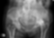
Medical brief of Legg-Calve-Perthes Disease, exploring the clinical signs, diagnostic evaluation, and long-term management of avascular necrosis in pediatric patients. FibonacciMD Compendium Pediatric Orthopedic Perthes Disease (Avascular Necrosis) Perthes Disease, more correctly named Legg Calve Perthes disease (LCPD) is a form of avascular necrosis of the capital femoral epiphysis in skeletally immature individuals. The disease can cause significant damage to the hip joint and can result in permanent disability. Treatment and Recommended Follow-Up Treatment of LCPD is complex depending upon the extent of the disease, age of the child, presence of epiphyseal collapse. It is recommended children with LCPD be referred to a competent pediatric orthopaedic surgeon. Treatment may involve physical therapy, casting, braces, and surgery. Diagnostic Evaluation and Differential Diagnosis Plain radographs are sufficient to establish the diagnosis. MRI may be somewhat helpful to predict the extent of the disease. Protein S & C should be evaluated if LCPD is bilateral. Signs and Symptoms The age of onset may be as young as 18 months, or as old as 9 years. The disease process usually runs a course of 2 years. In general the younger the child at the age on onset the better the prognosis. The earliest symptoms are a reduction of abduction of the affectd hip. Restricted internal rotation and a painless limp are often seen in LCPD. Radiographs may demonstrate widening of the joint space with or without the classic crescent subchondral fracture of the epiphysis early in the process. Pearl to Know Children with bilateral LCPD should have blood work to evaluate levels of Protien S and Protein C that regulate coagulation. These children may be predisposed to early myocardial infarction. FibonacciMD.app Compendium (our medical encyclopedia) has over 6,000 medical terms and medications.
- Aortic Valve Gradient and Critical Aortic Stenosis
Review aortic valve gradient staging & critical aortic stenosis criteria. Learn diagnostic benchmarks for mean gradients, valve area, and low-gradient AS staging. PART 1: Aortic Valve Gradient PART 2: Critical Aortic Stenosis FibonacciMD Compendium PART 1: Aortic Valve Gradient by Warren Rosenblum, M.D. Aortic Valve Gradient The aortic valve gradient is the difference in pressure between the left ventricle and the aortic root just distal to the aortic valve. Diagnostic Evaluation The aortic valve gradient can be obtained invasively during cardiac catheterization by measuring pressures directly from the left ventricle and the ascending aorta or estimated noninvasively using echocardiography. Staging An elevated aortic valve gradient suggests aortic stenosis. If the elevated gradient is found via echocardiography, correlation with aortic valve morphology is readily available. If the elevated aortic valve gradient is obtained at cardiac catheterization, echocardiography is appropriate to evaluate the aortic valve morphology. A mildly elevated gradient is < 20 mmHg, a moderately elevated gradient is 20-40 mmHg, and a severely elevated gradient is > 40 mmHg.1 Reference [1] Classification of valve stenosis and regurgitation. ECHOpedia Web site. http://www.echopedia.org/wiki/Classification_of_valve_stenosis_and_regurgitation. Updated December 11, 2016. Accessed May 15, 2017. Critical Aortic Stenosis by Richard Strongwater Critical aortic stenosis involves severe narrowing of the aortic valve orifice secondary to calcific aortic valve sclerosis, rheumatic heart disease, or congenital malformation. The mean gradient > 40 mmHg, and the valve area < 1.0 cm2. Causes and Risk Factors Critical aortic stenosis is related to congenital-unicuspid or bicuspid valve, aortic valve calcification, and rheumatic fever. Signs and Symptoms Symptoms of critical aortic stenosis include shortness of breath, decreased exercise tolerance, chest pain/pressure, near syncope, and, occasionally, congestive heart failure. Symptoms are often subtle as patients reduce physical activity with disease progression. Diagnostic Evaluation Transthoracic echocardiographic diagnosis of critical aortic stenosis is based on American College of Cardiology/American Heart Association guidelines for severe stenosis, including jet velocity > 4.0 m/sec, mean gradient > 40 mmHg, valve area < 1.0 cm2, valve area index < 0.6 cm/m2, and confirmation during cardiac catheterization with direct pressure measurements and calculated aortic valve area using Gorlin formula. Pearl Exercise testing may be warranted in "asymptomatic patients" to elicit left ventricular dysfunction or ischemia. Low-Gradient Aortic Stenosis Up to 40% of patients being considered for aortic valve replacement might have an aortic valve area ≤ 1 cm2 (indicating severe AS) but with only a moderate gradient: >20 and <40 mmHg (typical of moderate stenosis). Nevertheless, this would also be considered severe aortic stenosis and is known as low-gradient AS. The prognostic implications of low-gradient AS are mixed. However, the low-flow low-gradient AS with reduced left ventricular ejection fraction would have the worst prognosis and thus would also be considered critical AS. Treatment Surgical aortic valve replacement (bioprosthesis vs mechanical) or transcatheter aortic valve replacement (TAVR) may be indicated. FibonacciMD.app Compendium (our medical encyclopedia) has over 6,000 medical terms and medications. References Nishimura RA, Otto CM, Bonow RO, et al. 2014 AHA/ACC guideline for the management of patients with valvular heart disease: a report of the American College of Cardiology/American Heart Association Task Force on Practice Guidelines. Circulation. 2014;129:2440-2492. Roberts WC. Anatomically isolated aortic valvular disease: the case against its being of rheumatic etiology. Am J Med. 1970;49:151–159. Lewin MB, Otto CM. The bicuspid aortic valve: adverse outcomes from infancy to old age. Circulation. 2005;111:832-834. Rizzello V. Moderate gradient severe aortic stenosis: diagnosis, prognosis and therapy. Eur Heart J Suppl. 2021 Oct 8;23(Suppl E):E133-E137. doi: 10.1093/eurheartj/suab108. PMID: 34650372; PMCID: PMC8503314.
- Bicuspid Aortic Valve (BAV): Symptoms, Diagnosis, and Management
A Brief Review of BAV: The Most Common Congenital Heart Disease, Affecting 0.9%-1.36% of the Population FibonacciMD Compendium Bicuspid Aortic Valve (BAV) by Warren Rosenblum, MD Bicuspid aortic valve (BAV) involves the presence of only two leaflets in the aortic valve apparatus instead of the normal three leaflets. In this congenital condition, two leaflets are fused to create one. It is the most common congenital heart disease and is found in 0.9%-1.36% of the population. Most commonly, BAV involves fusion of the right coronary and left coronary cusps and, less often, of the right and noncoronary cusps. Fusion is associated with a dilated aortic root of the ascending aorta, aorticaneurysms, and aortic dissections. Over time, the valve may become stenotic and cause symptoms including congestive heart failure, hypertension, and syncope. Alternatively, BAV may result in regurgitation if the leaflets do not coapt correctly to cause dilation of the left ventricle. Patients with BAV also are at risk of developing endocarditis. Diagnostic Evaluation A bicuspid aortic valve often is discovered on physical exam. Auscultation of a systolic murmur best heard at the second right intercostal space is noted, prompting an echocardiographic examination. Confirmation can be obtained with transesophageal echocardiography or cardiac catheterization. It also may be evaluated by cardiac magnetic resonance imaging (MRI) or computed tomography (CT). Treatment and Recommended Follow-up Many patients survive long term without intervention. A significant percentage of patients require surgical repair if the valve is regurgitant or replacement if stenosis is present. If the aortic root is dilated or an aneurysm is found, the aorta may require repair or replacement. Asymptomatic patients < 30 years of age who have low Doppler gradients require echocardiographic follow-up every 1-2 years; those who have elevated AV Doppler gradients require yearly echocardiographic follow-up. Older adults with aortic stenosis or regurgitation require follow-up based on the degree of stenosis or regurgitation; it may vary between every 6 months if severe, 1-2 years if moderate, and 3-5 years if mild. Adults with an aortic root or ascending aorta > 4 cm should have a baseline CT or MRI; those with an aortic root or ascending aorta > 4.5 cm or who have a rapid change in aortic diameter should be imaged annually. FibonacciMD.app Compendium (our medical encyclopedia) has over 6,000 medical terms and medications. References Nishimura RA, Otto CM, Bonow RO, et al. 2014 AHA/ACC guideline for the management of patients with valvular heart disease: a report of the American College of Cardiology/American Heart Association Task Force on Practice Guidelines. Circulation. 2014;129:2440-2492. Roberts WC. Anatomically isolated aortic valvular disease: the case against its being of rheumatic etiology. Am J Med. 1970;49:151–159. Lewin MB, Otto CM. The bicuspid aortic valve: adverse outcomes from infancy to old age. Circulation. 2005;111:832-834.
- TAVR vs. SAVR: Comparing Approaches to Aortic Valve Replacement
Aortic Valve Replacement (AVR) and Transcatheter aortic-valve replacement (TAVR) Part 1: Aortic Valve Replacement Part 2: Transcatheter Aortic-Valve Replacement Cardiology PART 1: Aortic Valve Replacement by Cheng-Hung Tai, MD and David B. Grossberg, MD, FACC Aortic valve replacement (AVR) is a surgical procedure that is indicated for severe aortic stenosis(AS). Severe aortic stenosis is defined with a maximum aortic transvalvular velocity ≥4 m/sec and an aortic valve area ≤1 cm2. There are multiple treatment options, including: medical optimization which is usually of limited benefit at this grade of valvular stenosis, percutaneous balloon aortic valvotomy which is mostly a palliative treatment with only short-term benefits and high procedural risks and periprocedural CVA risks, transcatheter aortic valve replacement (TAVR) and surgical aortic valve replacement (SAVR). The choice of intervention will depend on patient's anticipated life expectancy and functional status. Indications A diagnosis of severe aortic stenosis can be made on physical examination but always necessitates a transthoracic echocardiography. Next, a heart valve replacement team should be consulted to assess the patient. If the patient's life expectancy is felt to be greater than 1 year if a surgical aortic valve replacement or transcatheter aortic valve replacement is performed AND if it is expected that the SAVR/TAVR procedure will improve the patient's quality of life, then the patient can be surgically risk assessed for TAVR or SAVR. Patients that are low to intermediate surgical risk with an inaccessible transfemoral TAVR (or any other adverse anatomic features) are recommended to undergo SAVR. Extreme and high surgical risk patients with an accessible transfemoral approach for TAVR are recommended to undergo TAVR. If this is not possible, then a careful risk/benefit assessment should be done to look for alternative sites for access for TAVR, or have the patient's medical therapy optimized. Current AHA/ACC guidelines for SAVR in patients with AS are as follows: Symptomatic severe high gradient AS Asymptomatic severe AS with left ventricular ejection fraction (LVEF) <50% Severe AS when undergoing coronary bypass or other cardiac surgery The latter two are strong considerations for valve replacement given risk for rapid disease progression: Asymptomatic very severe AS and low surgical risk Asymptomatic severe AS with decrease in systemic blood pressure or exercise tolerance during physical activity. The recently published SURTAVI trial showed TAVR is non-inferior to SAVR in a large randomized cohort of intermediate risk patients. (1) Primary endpoint: All-cause mortality or disabling stroke for TAVR vs. SAVR at 24 months: 12.6% vs. 14.0%, p < 0.05 for non-inferiority Coronary Revascularization Results : After randomization to treatment, there were 169 TAVR and PCI patients, 163 SAVR and coronary artery bypass grafting (CABG) patients, 695 TAVR only patients, and 633 SAVR only patients. Complete revascularization was not significantly different between TAVR + PCI vs. SAVR and CABG. No significant difference in the rate of the primary endpoint was found between TAVR and PCI, and SAVR and CABG. Overall Results : The results of this landmark trial indicate that TAVR is non-inferior to SAVR for the primary endpoint of mortality/disabling stroke at 24 months for the treatment of severe symptomatic aortic stenosis in intermediate-risk patients. Vascular complications were higher in TAVR patients at 30 days (6.0% vs.1.1% with SAVR). In the patients getting SAVR some complications were more frequent: new-onset atrial fibrillation was higher in the SAVR group (43% vs.12.9% with TAVR), as was the incidence of acute kidney injury, and the need for blood transfusions. 25.9% of patients required a new pacemaker post-TAVR, compared with 6.6% post-SAVR. Valve performance at 2 years was similar between the two strategies. From the patient’s perspective, TAVR provided earlier symptomatic benefit, but at 2 years of follow-up, symptoms were similar in both the TAVR and SAVR groups. Functional status improvement was also faster among patients undergoing TAVR rather than SAVR, but functional status measurements were similar in both groups at both 1 and 2 years of follow-up. On 5-year follow-up, clinical outcomes were similar. There were more repeat valve interventions post-TAVR, but the majority were within the first 2 years. This is a landmark trial in this field. Longer-term follow-up is going to be essential to understand long-term performance and the risk of other complications such as sub-clinical leaflet thrombosis which may lead to increased embolic events, and structural valve degeneration which can lead to increased risk of aortic insufficiency (AI). Sources and further reading: Nishimura RA, Otto CM, Bonow RO, et al. 2017 AHA/ACC Focused Update of the 2014 AHA/ACC Guideline for the Management of Patients With Valvular Heart Disease: A Report of the American College of Cardiology/American Heart Association Task Force on Clinical Practice Guidelines. J Am Coll Cardiol. 2017;70(2):252-289. Vahanian A, Alfieri O, Andreotti F, et al. Guidelines on the management of valvular heart disease (version 2012). Eur Heart J. 2012;33(19):2451-96. Goel SS, Agarwal S, Tuzcu EM, et al. Percutaneous coronary intervention in patients with severe aortic stenosis: implications for transcatheter aortic valve replacement. Circulation. 2012;125(8):1005-13. (1) Tuttle MK, Kiaii B, Van Mieghem NM, et al. Functional Status After Transcatheter and Surgical Aortic Valve Replacement: 2-Year Analysis From the SURTAVI Trial. JACC Cardiovasc Interv 2022;15:728-38. Editorial Comment: Wood DA, Sathananthan J. The Next Frontier in TAVR: Identifying Nonresponders and Treating Concomitant Pathologies. JACC Cardiovasc Interv 2022;15:728-38. PART 2: Transcatheter Aortic-Valve Replacement by Andrew Kupersmith, M.D. Transcatheter aortic-valve replacement (TAVR) often is performed in a manner similar to conventional cardiac catheterization. A folded balloon-mounted bioprosthetic valve is inserted via a femoral artery, advanced across the diseased aortic valve (stenotic, insufficient, or both), and then deployed by inflating the balloon. If a patient does not have femoral arteries of sufficient caliber, TAVR may be performed via a surgical approach through a small left lateral chest incision and then advanced through the left ventricular apex and across the native aortic valve. This obviates the need for a full sternotomy. Before being considered for a TAVR procedure, a patient must be evaluated at a center that performs the procedures frequently. Pear to Know TAVR procedures are recommended, and currently are only commercially approved, for patients who are poor candidates for standard surgical-valve replacements. Such patients tend to be older and too frail to tolerate a sternotomy procedure involving general anesthesia. Ongoing studies are evaluating the efficacy of TAVR procedures in healthier people. Author: Andrew Kupersmith Editor’s Comment - “TAVR may be performed via a surgical approach through a small left lateral chest incision”. In the early days of TAVR, the chest wall approach was anterolateral. I suspect that this approach in women may have seen a significant incidence of chest wall infections including MRSA. More medical terms in FibonacciMD.app Compendium (our medical encyclopedia).
- Pulmonary Valve Regurgitation: Clinical Diagnosis, Severity Criteria, and Management
Pulmonary valve regurgitation causes backflow through the pulmonic valve. Learn key signs including Graham Steell murmur, rSR EKG pattern, and AHA/ACC treatment guidance. FibonacciMD Compendium Pulmonary Valve Regurgitation by Christabel Nyange, MD, MPH and edited by Todd Pulerwitz MD Pulmonary valve regurgitation, also called pulmonary regurgitation (PR), pulmonic regurgitation or leaky pulmonary valve occurs when the pulmonic valve fails to close completely during diastole or for other reasons, allows blood to flow backwards into the right ventricle. Normal Heart Signs and Symptoms Most patients with pulmonary regurgitation are asymptomatic. However symptoms may include: exertional dyspnea - as a result of reduced cardiac output stemming from the volume overload to the right heart progressive decrease in exercise tolerance pedal edema, congestive hepatomegaly, and rarely, raised jugular venous distension (JVD) in severe PR symptomatic ventricular arrhythmia Physical findings in PR include: A diastolic decrescendo murmur heard in the 2nd and 3rd left intercostal spaces on auscultation A right-sided S3 could also be appreciated, indicating volume overload The pulmonary regurgitation murmur increases in intensity on inspiration due to the negative intrathoracic pressure generated (causing increased preload) and decreases in intensity with Valsalva maneuver Jugular venous distension with large v waves (Lancisi sign) can be seen in patients with PR secondary to pulmonary hypertension if the right ventricular pressure is high enough to cause severe tricuspid insufficiency as well The Graham Steell murmur: is a high pitched, blowing, early diastolic, decrescendo murmur (beginning immediately after P2), most prominent in the left 2 to 4 intercostal spaces, found with pulmonary hypertension. when there is dilatation of the pulmonary annulus causing high-velocity regurgitant jet. It increases in intensity with inspiration, exhibits little change after amyl nitrite inhalation or vasopressor administration, is diminished during the Valsalva strain, and returns to baseline intensity almost immediately after release of the Valsalva strain. Diagnostic Evaluation An echocardiogram is the investigation of choice for pulmonary regurgitation Echocardiogram frequently determines the severity, cause, mechanism, and determines progression to ventricular remodeling and heart failure. The severity of pulmonary regurgitation can be determined by a number of qualitative and semi-quantitative assessments, one of them is the width at the right ventricular outflow tract (RVOT) occupied by the regurgitant jet. It must be considered that eccentric jets may result in underestimation of PR jet severity. Mild PR - Normal right ventricular dimensions with narrow regurgitant jet width by color Doppler of < 25% of the pulmonary annulus Moderate PR - Normal or dilated right ventricle with intermediate regurgitant jet width (less than 50% of pulmonic valve annulus) Severe PR- Dilated right ventricle (except in acute PR) with large regurgitant jet width (greater than 50% of pulmonic valve annulus) Other investigations that can be used are: EKG: useful in patients who present with arrhythmias in the setting of right ventricular dilation secondary to volume overload. Commonly seen EKG findings include: rSR configuration in the right precordial leads (reflecting RV diastolic overload) in PR in the absence of pulmonary artery hypertension (PAH) P-pulmonale (tall p waves- indicating right atrial enlargement), increased r to s ratio in the right precordial leads along with right axis deviation (indicating RV hypertrophy), if PR is secondary to PAH Chest x-ray: lateral chest x-ray could reveal right ventricular dilation as reduced retrosternal space. Both pulmonary artery and right ventricular enlargement are visible, but these signs are nonspecific. Fluoroscopy may demonstrate pronounced pulsation of the main pulmonary artery. Cardiac MRI: can be used in moderate and severe PR. It helps in assessing pulmonary artery dilation and severity of PR by quantifying the regurgitant jet Angiography: varying degrees of opacification of the right ventricle are seen following the injection of contrast into the pulmonary artery due to the regurgitant jet. Causes and Risk Factors The most common cause of pulmonic regurgitation (PR) is the dilation of the valve annulus, however, it can be classified into physiologic, primary, and secondary causes. Physiological pulmonary regurgitation is considered an incidental finding on echocardiogram. Primary causes include: Iatrogenic: the most common causes are surgical valvotomy and balloon valvuloplasty infectious (infective endocarditis) with possible perforation or valve distortion Immune-mediated (rheumatic heart disease) Systemic (carcinoid disease) Congenital pulmonic regurgitation Post intervention: whether percutaneous or surgical valve replacement with valve failure or after operation or intervention for tetralogy of Fallot and valvular pulmonic stenosis Secondary or functional PR occurs in patients with a morphologically normal pulmonic valve who have: Idiopathic pulmonary artery dilatation and/or Pulmonary arterial hypertension, as a result of: Idiopathic Pulmonary arterial hypertension (PAH) Left heart disease (left heart failure, aortic and mitral valve disease) Lung disease (chronic obstructive lung disease, interstitial lung disease, obstructive sleep apnea) Chronic thromboembolism of lungs Other disorders of the pulmonary vasculature (sarcoidosis, sickle cell disease, schistosomiasis) Complications Untreated pulmonary regurgitation can result in: right ventricular dilation ventricular arrhythmias right heart failure congestive hepatomegaly anasarca left ventricular systolic dysfunction, and sudden cardiac death -- from ventricular arrhythmias Treatment Options Recommended treatment modalities as per the AHA/ACA guidelines are based on the severity of symptoms and the progression of PR. In asymptomatic patients with PR, treatment is not recommended unless 2/4 of the following criteria are met: mild or moderate RV or LV systolic dysfunction severe RV dilation (right ventricular end-diastolic volume >160ml/m2), right ventricular systolic pressure (RVSP) due to right ventricular outflow tract (RVOT) >2/3 of systemic pressure or progressively reduced exercise tolerance Symptomatic patients with PR can be considered for intervention -- whether by surgical pulmonic valve replacement with a bioprosthetic valve or now more frequently with a percutaneous pulmonic valve replacement (such as a Melody valve). Mechanical valves are rarely used. A bioprosthesis valve is preferred due to the durability of the prosthesis, low risk of valve failure, and no anticoagulation as compared to mechanical valves. Medical therapy is only recommended if the cause of pulmonary regurgitation is secondary to a primary medical condition such as pulmonary arterial hypertension or carcinoid disease or patient considered too high risk for interventional approach. In patients that are symptomatic with severe PR and are poor surgical candidates (due to age or comorbidities), medications targeting heart failure such as diuretics, ACEi/ARBs, and beta-blockers are recommended in appropriate situations. Sources and further reading: Corno AF. Pulmonary Valve Regurgitation: Neither Interventional Nor Surgery Fits All. Front Pediatr. 2018;6:169. Published 2018 Jun 7. doi:10.3389/fped.2018.00169 Saji AM, Sharma S. Pulmonary Regurgitation. [Updated 2020 Dec 14]. In: StatPearls [Internet]. Treasure Island (FL): StatPearls Publishing; 2020 Jan-. Available from: https://www.ncbi.nlm.nih.gov/books/NBK557564/ Pendela VS, Ayyad R. Pulmonic Regurgitation. [Updated 2020 May 6]. In: StatPearls [Internet]. Treasure Island (FL): StatPearls Publishing; 2020 Jan-. Available from: https://www.ncbi.nlm.nih.gov/books/NBK553136/